
Early Detection of Eye Diseases With Optomap
Early Detection of Eye Diseases With Optomap
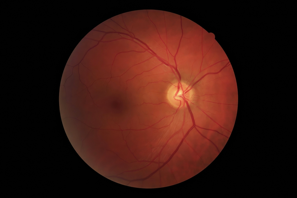
Some of the most serious eye diseases give no early warning. This is the reason early detection is so important. Ultra-widefield retinal imaging, such as Optomap, is helping eye doctors detect disease before it causes problems.
What Makes Optomap Different
A traditional eye exam only lets your doctor see a small part of your retina. To get a better look, your pupils usually need to be dilated. Even then, standard images show just a small area at the back of your eye.
Optomap is different. It uses a scanning laser to capture up to 200 degrees of your retina in one image. That’s about 82% of your retina at once, compared to only 15% with regular methods. The image is ready right away, so your doctor can review it with you during your visit.
This broader view is important because many diseases first show signs in the outer layers of the retina. Diabetic retinopathy is a good example. Studies show that up to half of diabetic lesions happen outside the area that a standard image would show. If you miss that part, you might miss early warning signs.
What Optomap Can Help Detect
Optomap imaging helps find many serious eye problems, often before you notice any symptoms. For example, it can detect diabetic retinopathy in its early stages. It does this by revealing lesions in the outer retina that might otherwise go unnoticed.
Glaucoma slowly damages the optic nerve. With Optomap, your doctor can monitor small changes over time by comparing year-to-year images. Macular degeneration can also be tracked this way. If small changes or spots appear in the outer retina, your doctor can intervene before your central vision is affected.
The far edges of the retina are where holes and tears often start. They may cause no pain or vision changes until they become serious. Optomap can spot them before they lead to detachment. It can also detect subtle changes associated with high blood pressure or diabetes. These conditions sometimes leave their earliest marks in the eyes.
What the Experience Is Like
An Optomap scan is fast and comfortable. You simply look into the device, and it takes the image in less than a second. There is no discomfort. Often, your pupils do not need to be dilated, so you will not have blurry vision or light sensitivity afterwards. You can drive yourself home and continue your day as usual.
The image is saved as part of your medical record. Each year, your doctor can compare it to earlier images and look for even small changes. This helps spot problems long before they affect your vision.
Why a Wider View Changes Things
Eye diseases are patient. They progress slowly. They do not always announce themselves with pain or blurred vision. That silence is what makes them dangerous.
A wider view of the retina gives your doctor a better chance of catching problems early. And early detection almost always leads to simpler treatment and better outcomes. It can mean the difference between managing a condition with minor interventions and facing permanent vision loss.
If you have diabetes, a family history of eye disease, or simply want the most thorough exam available, ask your eye doctor about Optomap imaging. A few seconds in front of a camera can protect your sight for years to come.
For more on early detection of eye diseases with optomap, visit SpecialEyes Eyecare. Our office is in Redmond, Washington. Call (425) 406-5397 to book an appointment today.
https://www.optos.com/blog/2025/august/diabetic-eye-disease-and-optomap-imaging/
https://www.atlantaeyegroup.com/ocular-disease-management.html

MAP

CONTACT INFORMATION
HOURS OF OPERATION
Monday 10:00am - 6:00pm
Tuesday 10:00am - 6:00pm
Wednesday 10:00am - 6:00pm
Thursday 10:00am - 6:00pm
Friday 10:00am - 6:00pm
Saturday 10:00am - 5:00pm
Sunday Closed
Powered by: 